Simulating fMRI Data
Bradley R. Buchsbaum
2026-09-25
Source:vignettes/a_08_simulation.Rmd
a_08_simulation.RmdIntroduction to fMRI Data Simulation
When checking an fMRI analysis pipeline, it is useful to have data
where the ground truth is known – real fMRI data do not come with known
effect sizes or noise parameters. Simulation supplies controlled
examples. The fmrireg package offers several functions with
varying levels of complexity:
-
simulate_bold_signal: Simulates clean BOLD responses for multiple experimental conditions -
simulate_noise_vector: Generates structured, fMRI-like noise with temporal autocorrelation, drift, and simplified physiological components -
simulate_simple_dataset: Combines signal and noise for a complete dataset based on SNR -
simulate_fmri_matrix: Creates multiple time series with shared event timing but column-specific variation in parameters
This vignette demonstrates their contracts and assumptions. The outputs are teaching simulations, not empirically validated generative models of scanner data.
Simulating Clean BOLD Signals
Let’s start with simulate_bold_signal, which generates a
clean hemodynamic response signal for multiple experimental
conditions.
# Simulate 3 conditions with different amplitudes
sim <- simulate_bold_signal(ncond = 3, amps = c(1.0, 1.5, 2.0), TR = 2)

# Extract the data
time <- sim$mat[,1]  # First column contains time
signals <- sim$mat[,-1]  # Other columns contain condition signals
We reshape the data into long format so ggplot2 can map each condition to a separate color.
df <- data.frame(
  Time = time,
  Cond1 = signals[,1],
  Cond2 = signals[,2],
  Cond3 = signals[,3]
)

df_long <- tidyr::pivot_longer(df, cols = c(Cond1, Cond2, Cond3),
                        names_to = "Condition",
                        values_to = "Response")
ggplot(df_long, aes(x = Time, y = Response, color = Condition)) +
  geom_line() +
  theme_minimal() +
  labs(title = "Simulated BOLD Signals for Three Conditions",
       x = "Time (seconds)",
       y = "BOLD Response",
       color = "Condition") +
  scale_color_brewer(palette = "Set1")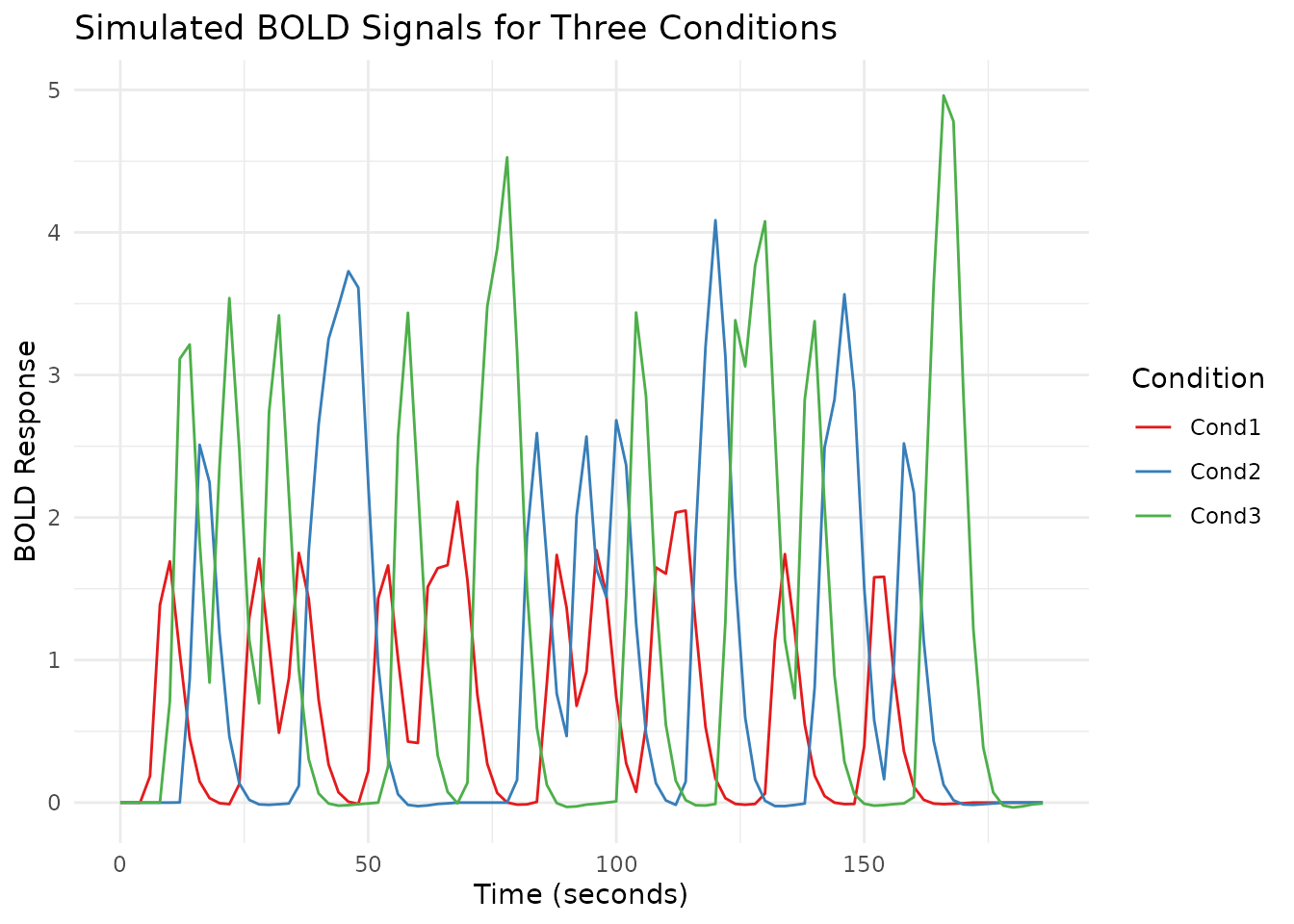
Adding dashed vertical lines at each event onset makes the relationship between stimulus timing and the hemodynamic response easier to see.
ggplot(df_long, aes(x = Time, y = Response, color = Condition)) +
  geom_line() +
  geom_vline(xintercept = sim$onset, linetype = "dashed", alpha = 0.3) +
  theme_minimal() +
  labs(title = "Simulated BOLD Signals with Event Onsets",
       x = "Time (seconds)",
       y = "BOLD Response",
       color = "Condition") +
  scale_color_brewer(palette = "Set1")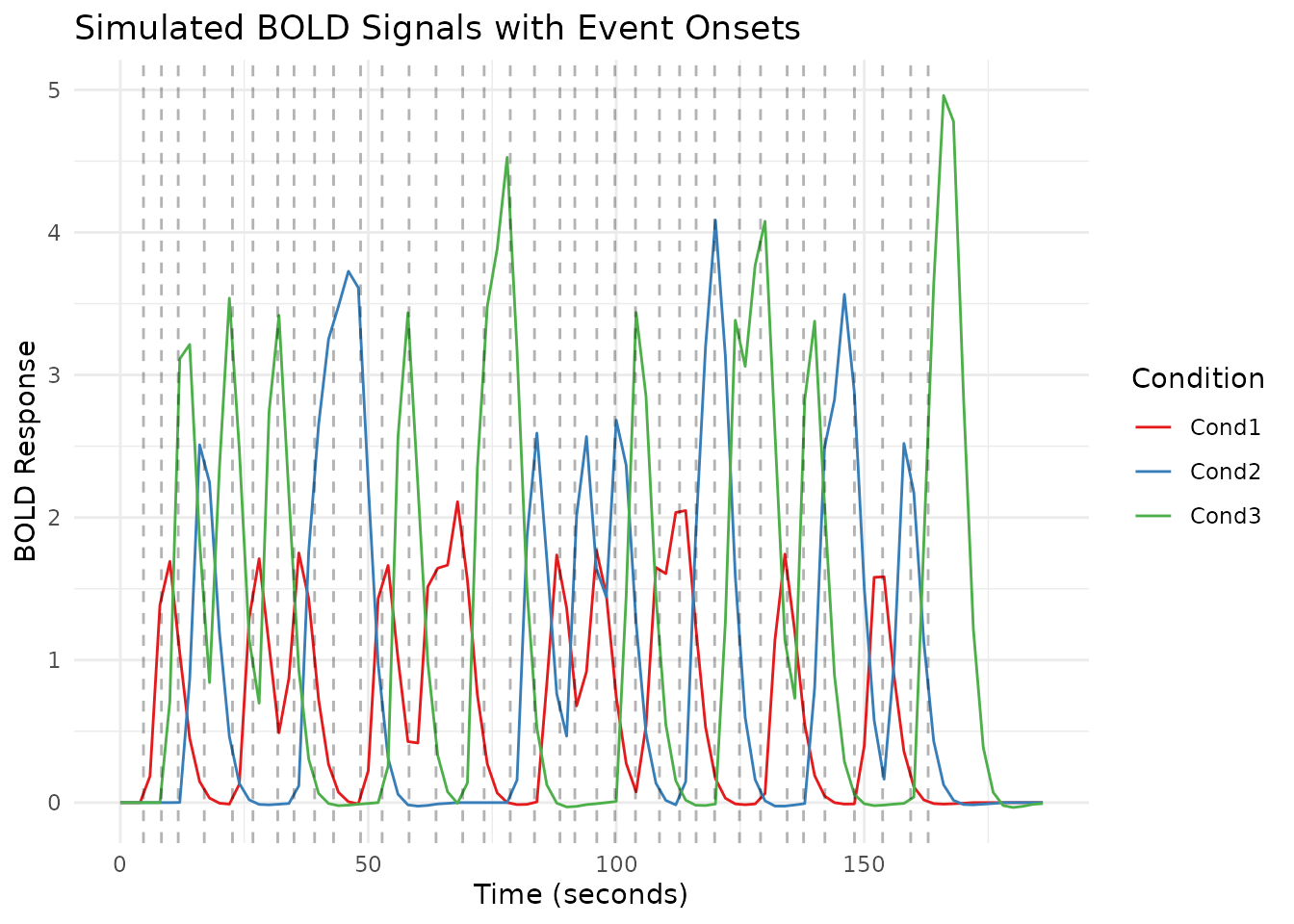
The function returns a list containing:
-
onset: Event onset times -
condition: Condition labels for each event -
mat: Matrix with time points and BOLD responses for each condition
You can control:
- Number of conditions (
ncond) - Number of repetitions per condition (
nreps) - HRF shape (
hrf) - Amplitudes for each condition (
amps) - Inter-stimulus interval range (
isi) - Amplitude variability (
ampsd)
Simulating Structured fMRI-like Noise
The simulate_noise_vector function combines stylized
components motivated by common fMRI noise sources. To understand exactly
what it generates, we simulate and visualize each component separately
and then combine them.
First, pure white noise – random fluctuations with no temporal structure.
noise_white <- simulate_noise_vector(n_timepoints, TR = TR,
                                  ar = numeric(0), ma = numeric(0),
                                  drift_amplitude = 0, physio = FALSE, sd = 1)
Next, ARMA noise introduces temporal autocorrelation, which makes the signal “smoother” than white noise.
noise_arma <- simulate_noise_vector(n_timepoints, TR = TR,
                                 ar = c(0.6), ma = c(0.3),
                                 drift_amplitude = 0, physio = FALSE, sd = 1)
Scanner drift is a slow oscillation that shifts the baseline over the course of a run.
drift_freq <- 1/128
drift_amplitude <- 2
noise_drift <- drift_amplitude * sin(2 * pi * drift_freq * time)
Physiological noise originates in continuous time, while an fMRI scanner records only one sample every TR. Here respiration is 0.3 Hz and cardiac activity is 1.1 Hz. Both exceed the 0.25 Hz Nyquist frequency at TR = 2 seconds, so they appear at lower frequencies after sampling: 0.2 Hz and 0.1 Hz, respectively. Generating the continuous signals first makes that aliasing explicit.
physio_time <- seq(0, max(time), by = 0.05)
cardiac_continuous <- 0.5 * sin(2 * pi * 1.1 * physio_time)
respiratory_continuous <- 0.8 * sin(2 * pi * 0.3 * physio_time)

noise_cardiac <- approx(physio_time, cardiac_continuous, xout = time)$y
noise_respiratory <- approx(physio_time, respiratory_continuous, xout = time)$y
noise_physio <- noise_cardiac + noise_respiratory
Finally, we combine the ARMA, drift, and sinusoidal physiological components into the declared structured teaching model. This sum illustrates separable noise sources; it is not an empirically validated scanner-noise distribution.
noise_combined <- noise_arma + noise_drift + noise_physio
noise_df <- data.frame(
  Time = time,
  White_Noise = noise_white,
  ARMA_Noise = noise_arma,
  Drift_Component = noise_drift,
  Physiological_Noise = noise_physio,
  Combined_Noise = noise_combined
)

noise_long <- tidyr::pivot_longer(noise_df,
                                  cols = -Time,
                                  names_to = "NoiseType",
                                  values_to = "Signal")

noise_long$NoiseType <- factor(noise_long$NoiseType,
                              levels = c("White_Noise", "ARMA_Noise",
                                        "Drift_Component", "Physiological_Noise",
                                        "Combined_Noise"),
                              labels = c("White Noise", "ARMA Noise",
                                        "Drift Component", "Physiological Noise",
                                        "Combined Noise"))
ggplot(noise_long, aes(x = Time, y = Signal, color = NoiseType)) +
  geom_line() +
  facet_wrap(~NoiseType, ncol = 1, scales = "free_y") +
  theme_minimal() +
  theme(legend.position = "none") +
  labs(title = "Simulated fMRI Noise Components: Time Courses",
       x = "Time (seconds)",
       y = "Signal Amplitude")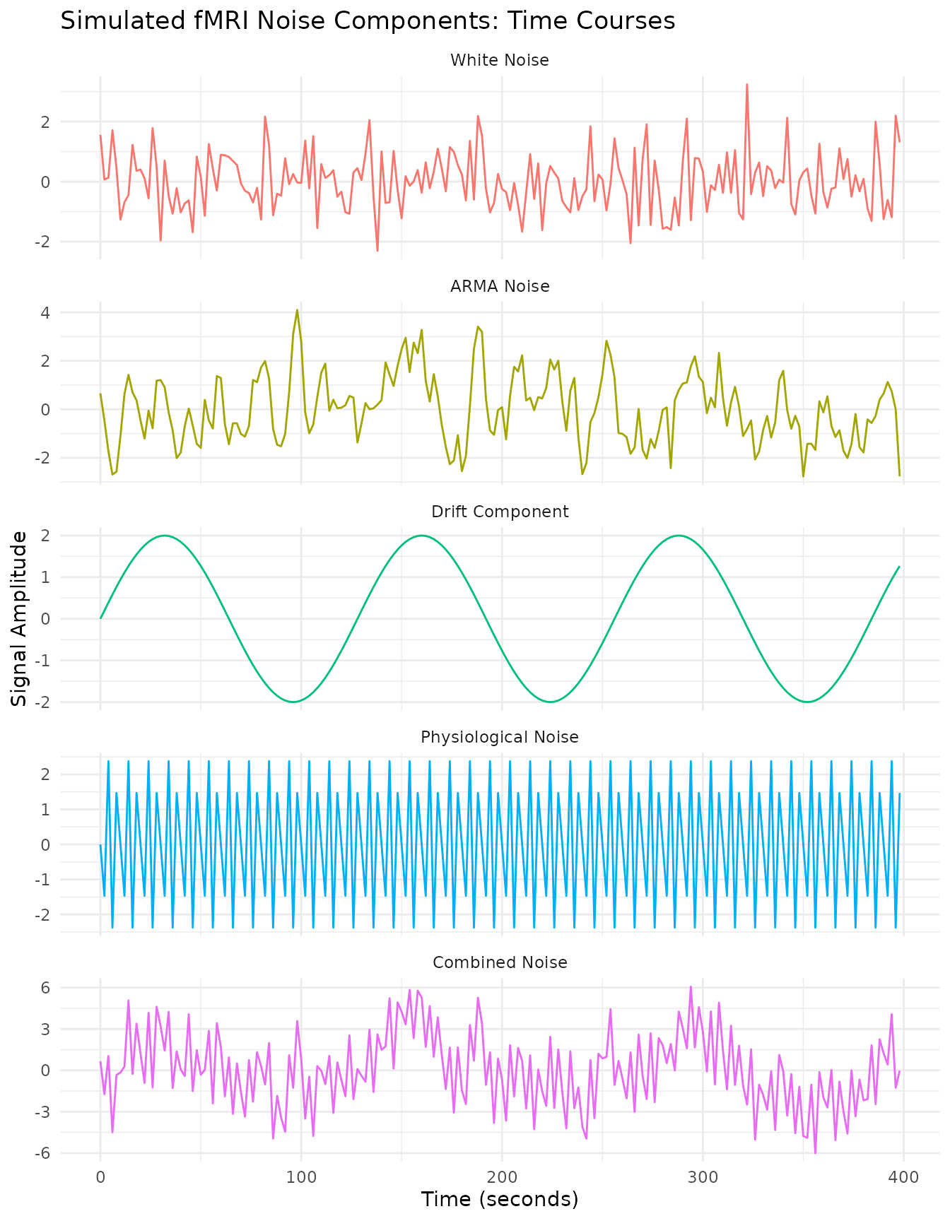
Power Spectrum Analysis
Power spectra reveal the frequency content of each noise type. White noise has a flat spectrum, while ARMA noise concentrates power at lower frequencies.
spec_white <- calculate_spectrum(noise_white, TR)
spec_arma <- calculate_spectrum(noise_arma, TR)
spec_drift <- calculate_spectrum(noise_drift, TR)
spec_physio <- calculate_spectrum(noise_physio, TR)
spec_combined <- calculate_spectrum(noise_combined, TR)
spec_white$NoiseType <- "White Noise"
spec_arma$NoiseType <- "ARMA Noise"
spec_drift$NoiseType <- "Drift Component"
spec_physio$NoiseType <- "Physiological Noise"
spec_combined$NoiseType <- "Combined Noise"

spec_df <- rbind(spec_white, spec_arma, spec_drift, spec_physio, spec_combined)

spec_df$NoiseType <- factor(spec_df$NoiseType,
                           levels = c("White Noise", "ARMA Noise",
                                     "Drift Component", "Physiological Noise",
                                     "Combined Noise"))
We also compute a high-resolution drift spectrum from a longer signal to better resolve the low-frequency peak.
n_long <- 1024
time_long <- seq(0, (n_long - 1) * TR, by = TR)
drift_long <- drift_amplitude * sin(2 * pi * drift_freq * time_long)
spec_drift_long <- calculate_spectrum(drift_long, TR)
spec_drift_long$NoiseType <- factor(
  "Drift Component",
  levels = levels(spec_df$NoiseType)
)
Each panel below shows the power spectrum of a different noise component. The dashed green line in the drift panel shows the high-resolution version.
ggplot() +
  geom_line(data = spec_df, aes(x = Frequency, y = Power, color = NoiseType)) +
  geom_line(data = subset(spec_drift_long, Frequency <= 0.05),
           aes(x = Frequency, y = Power), color = "darkgreen", linetype = "dashed") +
  theme_minimal() +
  facet_wrap(~NoiseType, ncol = 1, scales = "free_y") +
  theme(legend.position = "none") +
  labs(title = "Power Spectra of Different Noise Components",
       x = "Frequency (Hz)",
       y = "Power") +
  scale_color_brewer(palette = "Set1") +
  coord_cartesian(xlim = c(0, 0.25))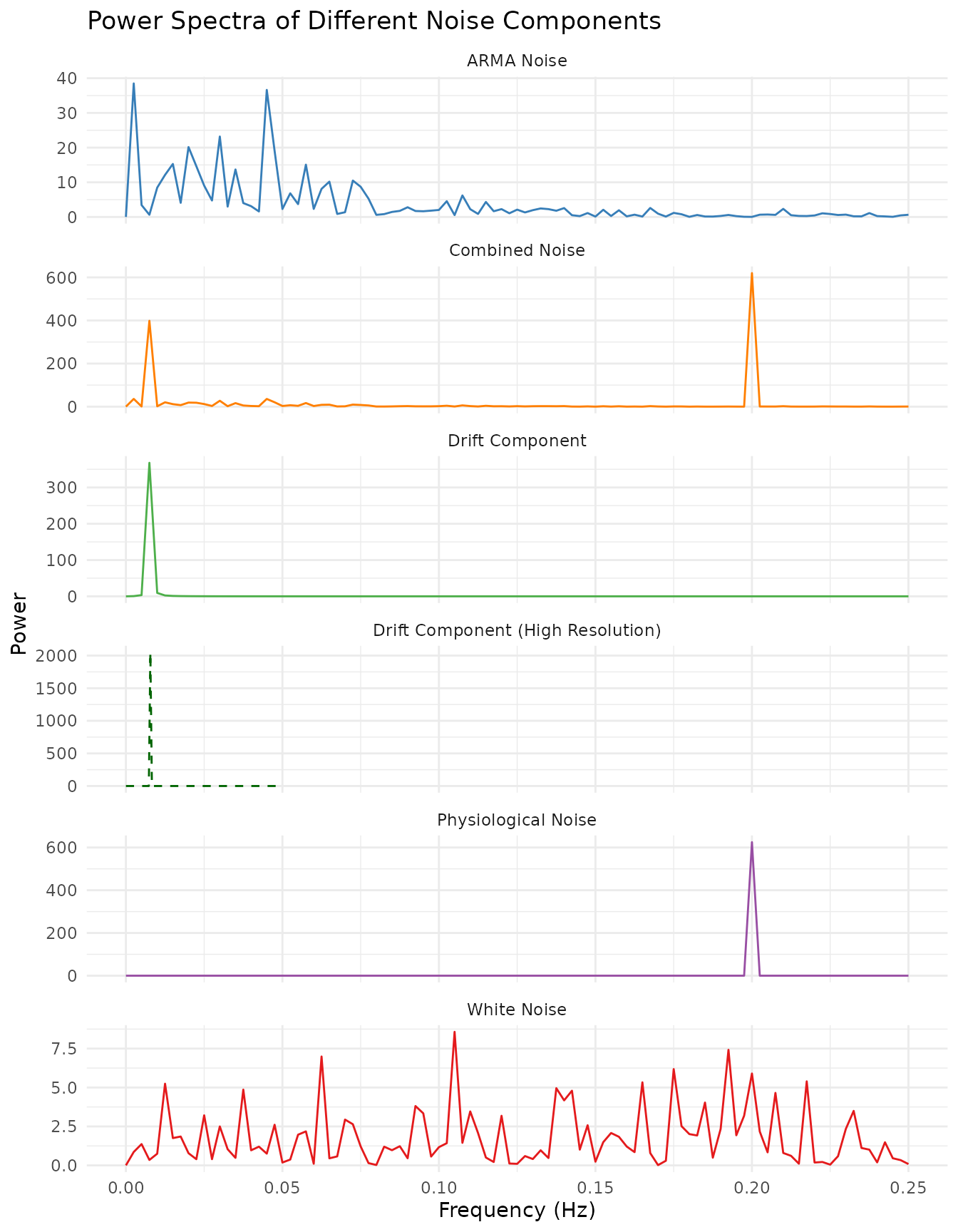
Plotting all spectra on a single log-scaled axis makes it easy to compare their relative magnitudes.
ggplot(spec_df, aes(x = Frequency, y = Power, color = NoiseType)) +
  geom_line() +
  theme_minimal() +
  labs(title = "Power Spectra of Different Noise Components (Log Scale)",
       x = "Frequency (Hz)",
       y = "Power (log scale)") +
  scale_y_log10() +
  scale_color_brewer(palette = "Set1") +
  guides(color = guide_legend(nrow = 2, byrow = TRUE)) +
  theme(
    legend.position = "bottom",
    legend.title = element_blank(),
    legend.text = element_text(size = 8)
  ) +
  coord_cartesian(xlim = c(0, 0.25))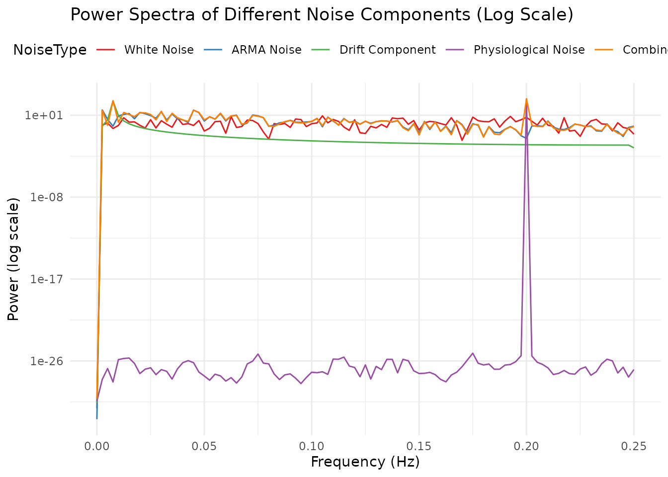
Zooming in on the very low-frequency region confirms the drift component peaks at the expected frequency (marked by the red dotted line).
drift_freq_idx <- which.min(abs(spec_df$Frequency - drift_freq))

ggplot() +
  geom_line(data = subset(spec_df, NoiseType == "Drift Component"),
           aes(x = Frequency, y = Power), color = "darkgreen") +
  geom_line(data = subset(spec_drift_long, Frequency <= 0.05),
           aes(x = Frequency, y = Power), color = "darkgreen", linetype = "dashed") +
  geom_vline(xintercept = drift_freq, linetype = "dotted", color = "red") +
  theme_minimal() +
  labs(title = "Zoomed Power Spectrum of Drift Component",
       subtitle = paste("Expected peak at", drift_freq, "Hz"),
       x = "Frequency (Hz)",
       y = "Power") +
  coord_cartesian(xlim = c(0, 0.05))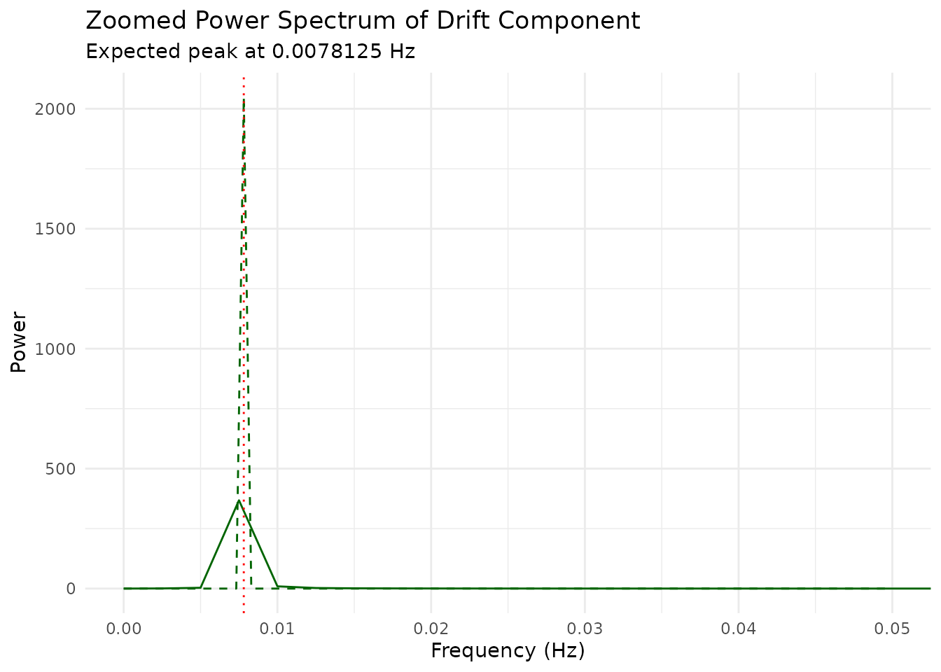
The simulation shows five distinct types of noise components:
White Noise: Random fluctuations with equal power across all frequencies (flat power spectrum).
ARMA Noise: Temporally autocorrelated noise with a characteristic “smoothed” appearance. The power spectrum shows more power at lower frequencies.
Drift Component: A very slow oscillation typical of scanner drift or physiological trends. The power spectrum shows a dominant peak at a very low frequency.
Physiological Noise: Simplified respiratory and cardiac sinusoids at 0.3 Hz and 1.1 Hz are above the 0.25 Hz Nyquist limit. At TR = 2 seconds they appear at distinct aliases of 0.2 Hz and 0.1 Hz. These components demonstrate sampling and aliasing; they are not a validated physiological-noise model, nor evidence that the underlying physiology occurs at the aliased frequencies.
Combined Noise: The sum of the stylized components. Its power spectrum shows features from each contributor.
Adding these components to task-related signals creates controlled, structured time series for demonstrations and numerical checks.
Creating a Complete Dataset with Signal and Noise
The simulate_simple_dataset function combines clean
signals and noise to create a complete fMRI dataset with a specified
signal-to-noise ratio (SNR).
snr_values <- c(1.0, 0.5, 0.2)
snr_datasets <- lapply(snr_values, function(snr) {
  simulate_simple_dataset(ncond = 3, TR = 2, snr = snr, seed = 42)
})
names(snr_datasets) <- paste0("SNR = ", snr_values)

data_snr_1.0 <- snr_datasets[["SNR = 1"]]
data_snr_0.5 <- snr_datasets[["SNR = 0.5"]]
data_snr_0.2 <- snr_datasets[["SNR = 0.2"]]
The same seed deliberately preserves the event schedule, clean signal, and standardized noise realization. Only the scale of that noise changes, so the panels isolate SNR rather than comparing three unrelated simulations.
plot_df <- rbind(
  create_plot_df(data_snr_1.0, "SNR = 1.0"),
  create_plot_df(data_snr_0.5, "SNR = 0.5"),
  create_plot_df(data_snr_0.2, "SNR = 0.2")
)

plot_df_long <- tidyr::pivot_longer(plot_df,
                                    cols = -c(Time, SNR),
                                    names_to = "Type",
                                    values_to = "Signal")
The overlay plot shows the clean signal (red) against the noisy measurement (blue) at each SNR level. As SNR decreases, the noise increasingly obscures the true signal.
ggplot(plot_df_long, aes(x = Time, y = Signal, color = Type)) +
  geom_line() +
  facet_wrap(~SNR, ncol = 1) +
  theme_minimal() +
  labs(title = "Clean vs. Noisy Signals at Different SNR Levels",
       subtitle = "Single condition shown for clarity",
       x = "Time (seconds)",
       y = "Signal",
       color = "Signal Type") +
  scale_color_manual(values = c("Clean" = "darkred", "Noisy" = "steelblue")) +
  theme(legend.position = "top")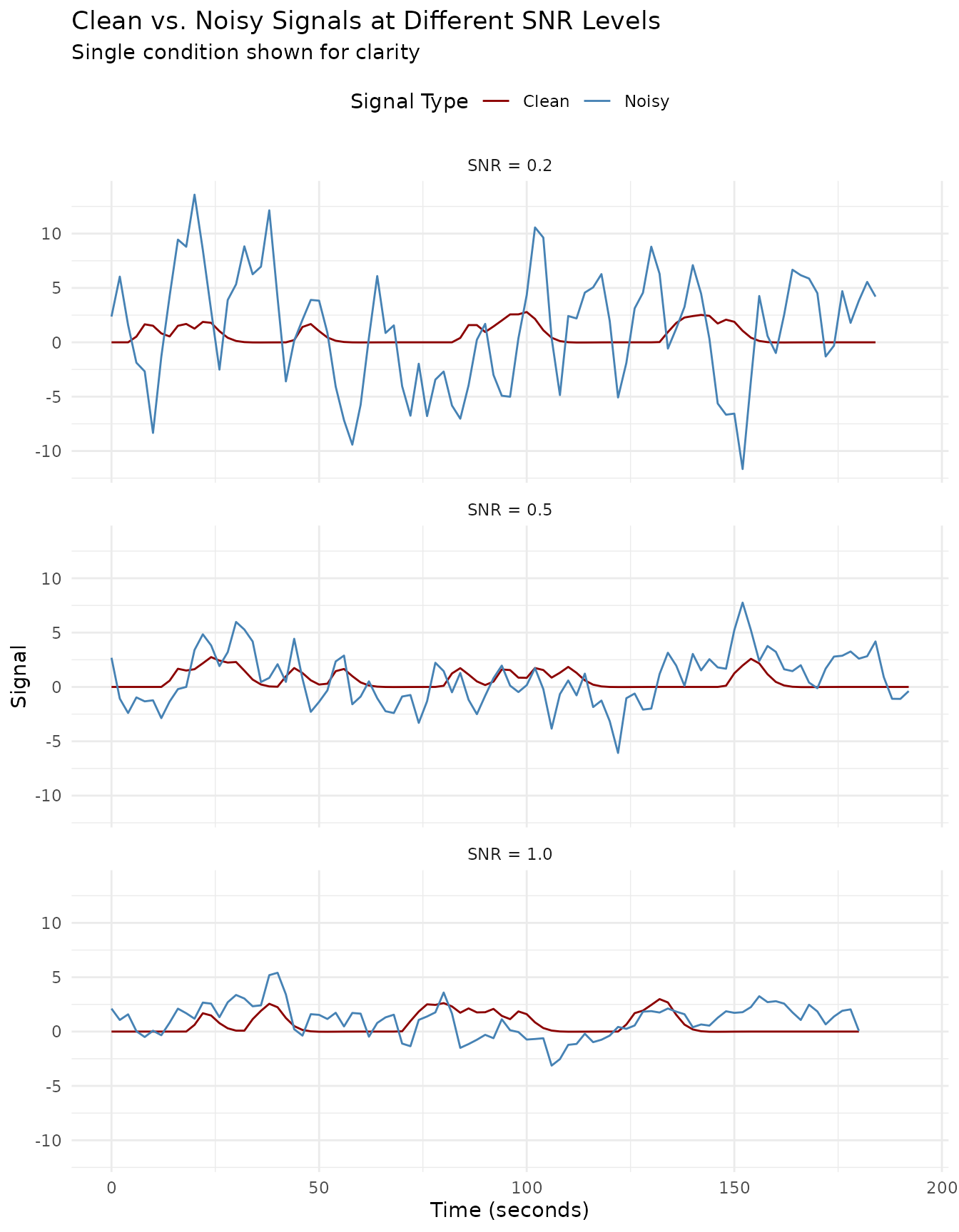
The decomposition plots below separate each SNR level into its signal, noise, and combined components. At SNR = 1.0 the signal is clearly visible; by SNR = 0.2 the noise dominates.
plot_faceted(plot_df, "SNR = 1.0")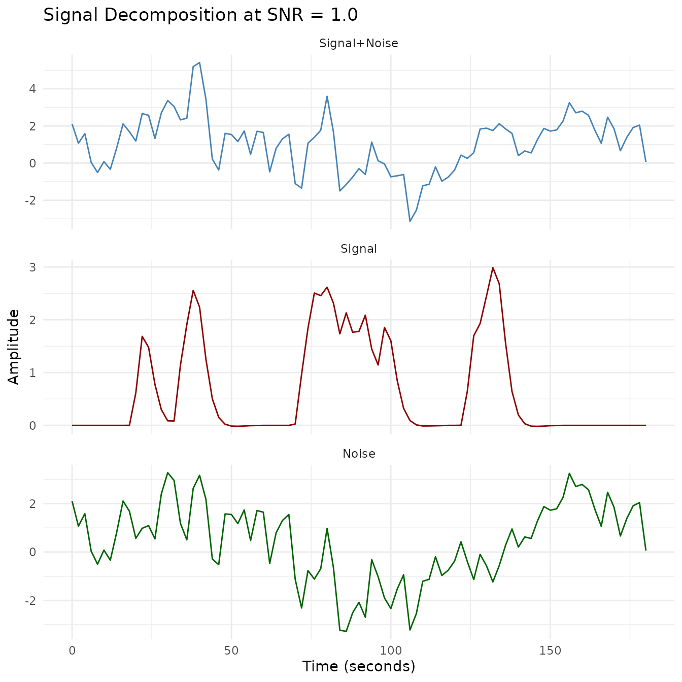
plot_faceted(plot_df, "SNR = 0.5")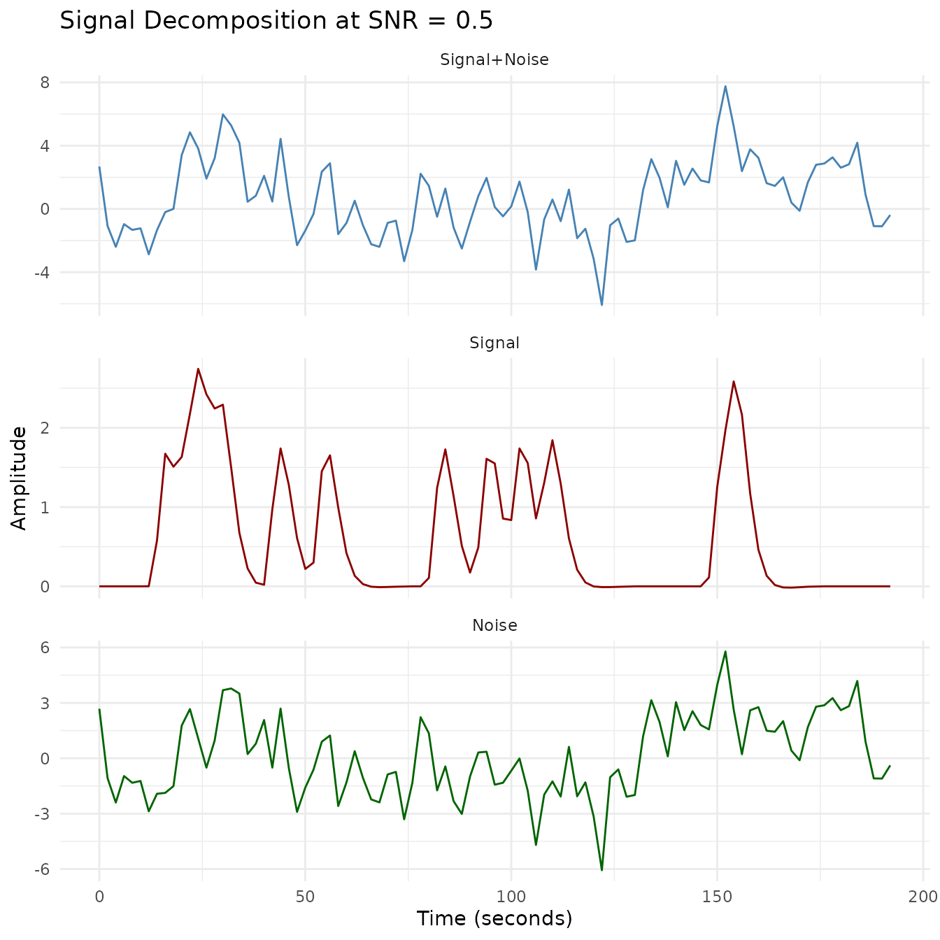
plot_faceted(plot_df, "SNR = 0.2")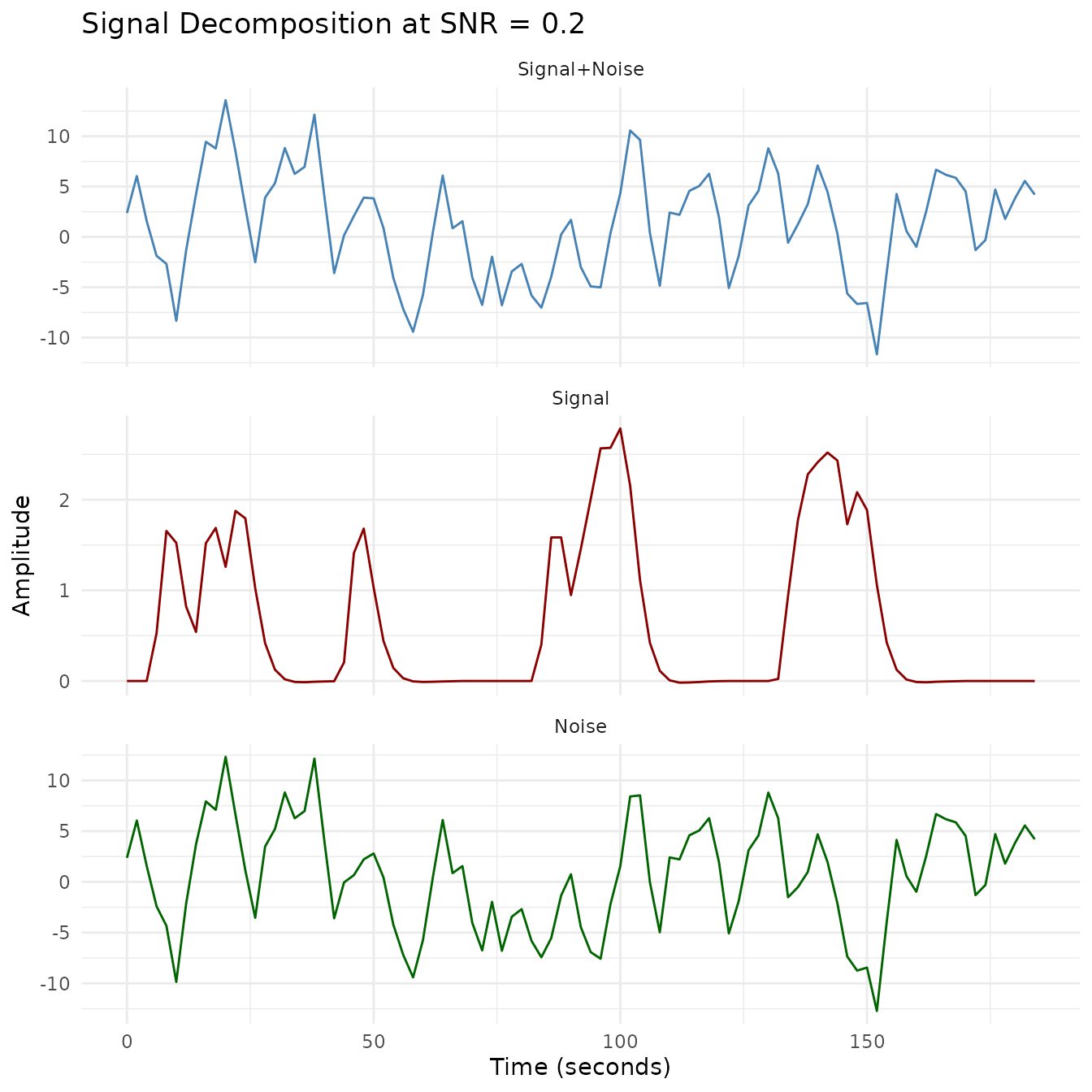
snr_stats <- data.frame(
  Requested_SNR = snr_values,
  Empirical_SNR = unname(empirical_snr),
  Signal_SD = vapply(snr_datasets, function(dataset) {
    sd(as.vector(dataset$clean$mat[, -1]))
  }, numeric(1)),
  Noise_SD = vapply(snr_datasets, function(dataset) {
    sd(as.vector(dataset$noise))
  }, numeric(1))
)
knitr::kable(
  snr_stats,
  digits = 3,
  caption = "Requested and empirically realized SNR from a controlled simulation"
)| Requested_SNR | Empirical_SNR | Signal_SD | Noise_SD | |
|---|---|---|---|---|
| SNR = 1 | 1.0 | 1.0 | 0.861 | 0.861 |
| SNR = 0.5 | 0.5 | 0.5 | 0.861 | 1.722 |
| SNR = 0.2 | 0.2 | 0.2 | 0.861 | 4.306 |
This visualization shows how different SNR levels affect the fMRI time series. The lower the SNR, the more the noise dominates the signal. For each SNR level, we show:
- The original clean signal (red line): The true underlying BOLD response
- The noisy signal (blue line): The synthetic observation produced by this model
- Signal decomposition: Visualization of how the signal, noise, and combined signal relate at each SNR level
With SNR = 1.0, the signal pattern remains visible despite the noise. At SNR = 0.5, some features are obscured; at SNR = 0.2, the noise substantially masks the signal in this controlled example.
The function returns:
-
clean: The simulated signals without noise -
noisy: The signals with added noise -
noise: The simulated noise component -
onsets: Trial onset times -
conditions: Condition labels for each trial
Simulating Matrix Time Series with Column-Specific Variation
The most advanced simulation function,
simulate_fmri_matrix, generates multiple time series
(columns) that share the same event timing but have column-specific
variations in amplitudes and durations. This is particularly useful for
simulating multiple voxels or regions with related but slightly
different response profiles.
requested_onsets <- seq(12, 156, length.out = 10)
sim_matrix <- simulate_fmri_matrix(
  n = 5,                  # 5 voxels/regions
  total_time = 200,       # 200 seconds of scan time
  TR = 2,                 # TR = 2 seconds
  onsets = requested_onsets,
  amplitudes = 1,         # Base amplitude = 1
  amplitude_sd = 0.3,     # Amplitude variability
  durations = 2,          # Base duration = 2 seconds
  duration_sd = 0.5,      # Duration variability
  noise_type = "ar1",     # AR(1) noise
  noise_sd = 0.5          # Noise standard deviation
)
When you supply onsets, those values are authoritative:
the function skips ISI generation and derives the event count from their
length.
ts_data <- sim_matrix$time_series
matrix_data <- fmridataset::collect_assay(ts_data)

time_points <- seq(0, by = 2, length.out = nrow(matrix_data))
plot_data <- data.frame(Time = time_points)

for(i in 1:ncol(matrix_data)) {
  plot_data[[paste0("Voxel", i)]] <- matrix_data[, i]
}

plot_data_long <- tidyr::pivot_longer(
  plot_data,
  cols = starts_with("Voxel"),
  names_to = "Voxel",
  values_to = "Signal"
)
All five simulated voxels share the same event timing, but their response amplitudes and durations vary independently.
ggplot(plot_data_long, aes(x = Time, y = Signal, color = Voxel)) +
  geom_line(alpha = 0.8) +
  theme_minimal() +
  labs(title = "Multiple Simulated Time Series with Column-Specific Variation",
       x = "Time (seconds)",
       y = "Signal",
       color = "Voxel") +
  scale_color_brewer(palette = "Set2")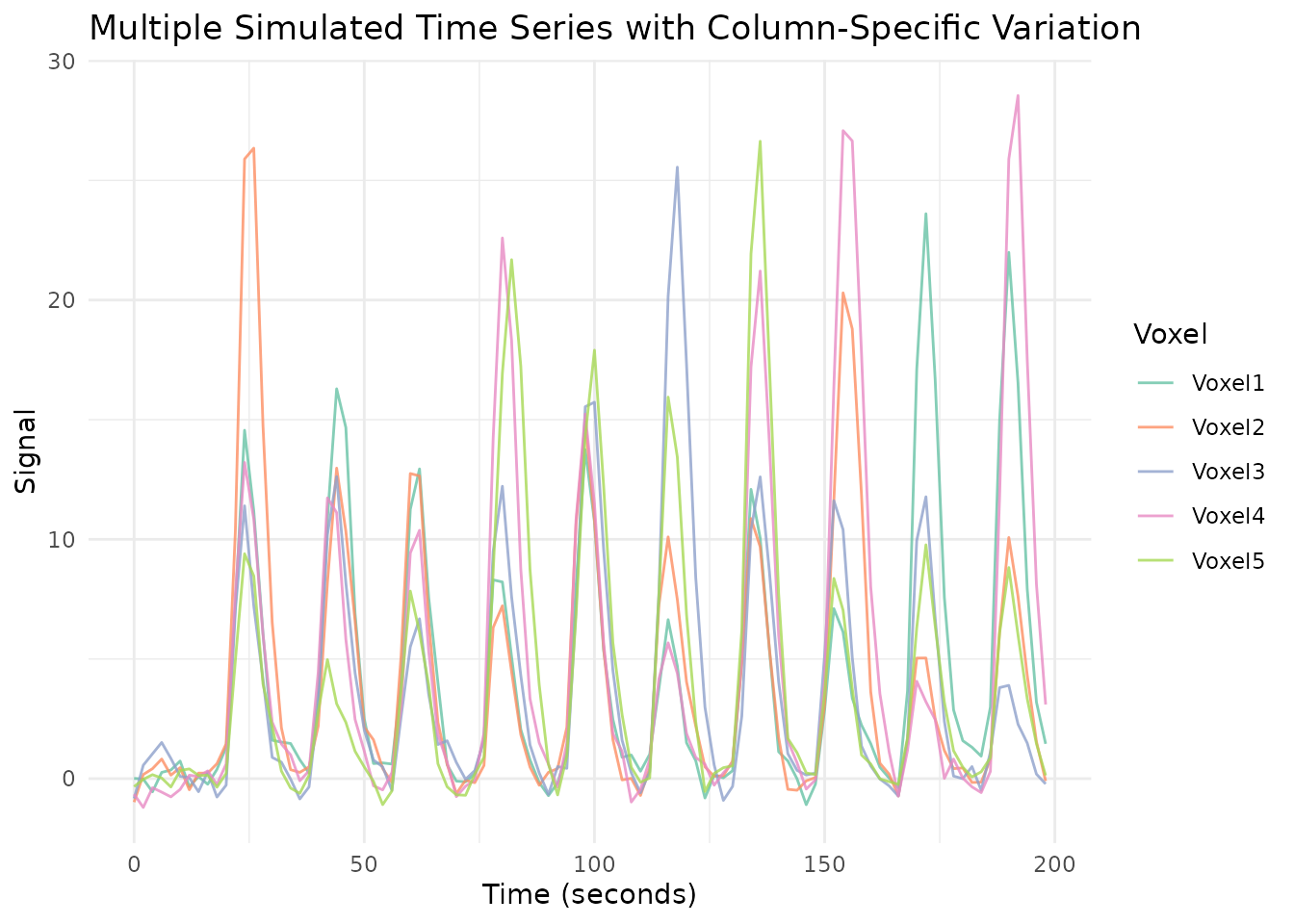
amp_df <- as.data.frame(sim_matrix$ampmat)
colnames(amp_df) <- paste0("Voxel", 1:ncol(amp_df))
amp_df$Event <- 1:nrow(amp_df)

dur_df <- as.data.frame(sim_matrix$durmat)
colnames(dur_df) <- paste0("Voxel", 1:ncol(dur_df))
dur_df$Event <- 1:nrow(dur_df)

amp_long <- tidyr::pivot_longer(
  amp_df,
  cols = starts_with("Voxel"),
  names_to = "Voxel",
  values_to = "Amplitude"
)

dur_long <- tidyr::pivot_longer(
  dur_df,
  cols = starts_with("Voxel"),
  names_to = "Voxel",
  values_to = "Duration"
)
The amplitude plot shows how each voxel’s response magnitude varies from event to event around the base amplitude of 1.
ggplot(amp_long, aes(x = Event, y = Amplitude, color = Voxel, group = Voxel)) +
  geom_line() +
  geom_point() +
  theme_minimal() +
  labs(title = "Amplitude Variation Across Events and Voxels",
       x = "Event Number",
       y = "Amplitude",
       color = "Voxel") +
  scale_color_brewer(palette = "Set2")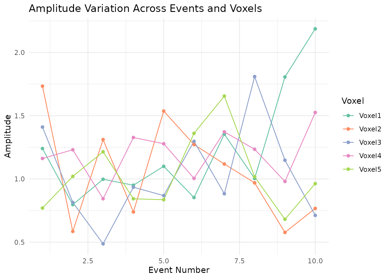
Duration variation follows the same pattern – each voxel draws its event durations independently around the base of 2 seconds.
ggplot(dur_long, aes(x = Event, y = Duration, color = Voxel, group = Voxel)) +
  geom_line() +
  geom_point() +
  theme_minimal() +
  labs(title = "Duration Variation Across Events and Voxels",
       x = "Event Number",
       y = "Duration (seconds)",
       color = "Voxel") +
  scale_color_brewer(palette = "Set2")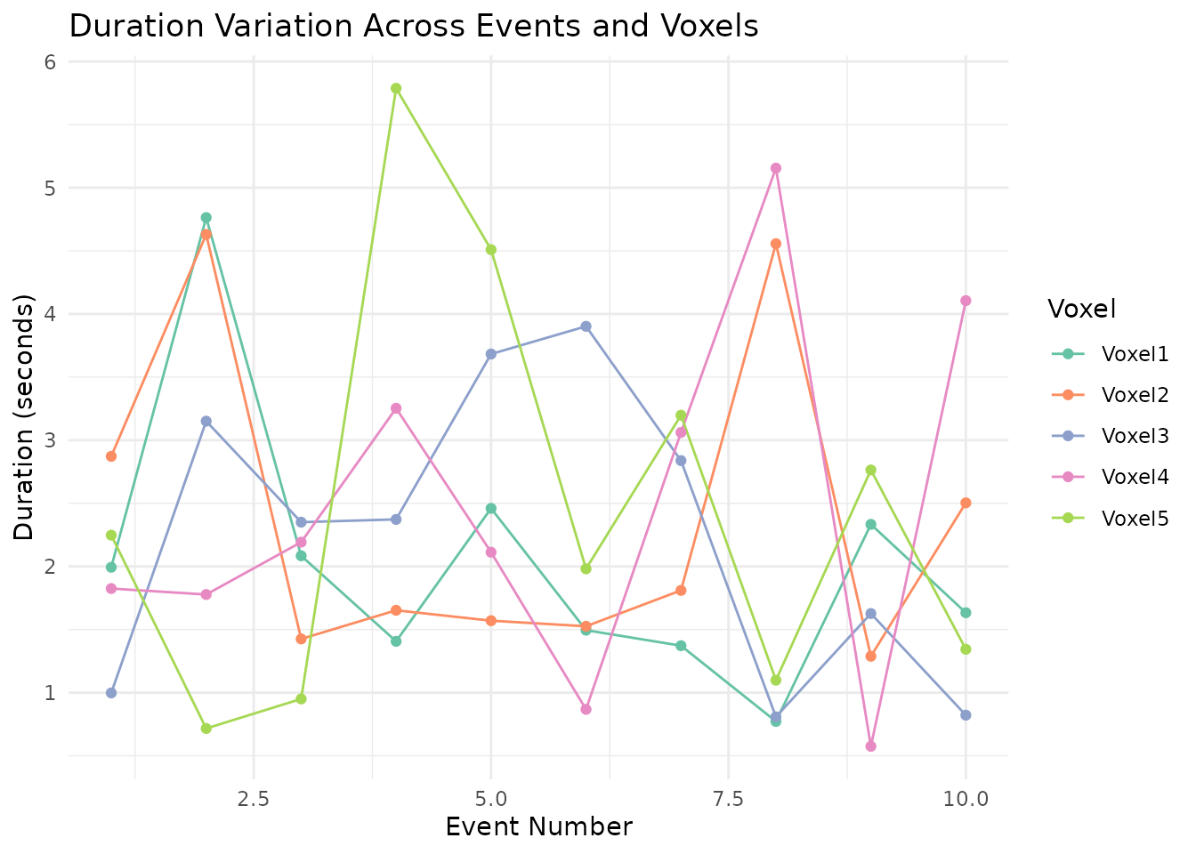
This function exposes the following controls for multiple related time series:
-
Shared event timing but individual variation in:
- Amplitude (per event, per column)
- Duration (per event, per column)
- Independent noise generation for each column
-
Complex output including:
- Time series matrix
- Amplitude and duration matrices
- HRF and noise parameter information
Summary and Comparison
The four simulation functions in fmrireg serve different
purposes and offer increasing levels of complexity:
-
simulate_bold_signal: Generate clean BOLD signals for multiple conditions -
simulate_noise_vector: Create structured, fMRI-like noise with temporal structure -
simulate_simple_dataset: Combine signal and noise with a specific SNR -
simulate_fmri_matrix: Create multiple time series with trial-by-trial, column-specific parameter variation
Choose the appropriate function based on your simulation needs:
- For basic signal generation: use
simulate_bold_signal - For structured teaching noise: use
simulate_noise_vector - For a complete dataset with controlled SNR: use
simulate_simple_dataset - For simulating multiple voxels/regions with shared timing but
response variation: use
simulate_fmri_matrix
These functions provide controlled inputs for implementation checks and teaching. Their explicit stochastic and signal assumptions should be inspected before using them as a benchmark for a scientific method.
Next
-
vignette("a_09_linear_model", package = "fmrireg")— Fit and interpret an fMRI linear model