Publication-quality continuous surface figures
neurosurf authors
2026-10-16
Source:vignettes/surface-figures.Rmd
surface-figures.RmdYou have a continuous value at every cortical vertex–for example a t, z, thickness, or connectivity map–and need a static figure that remains scientifically interpretable after thresholding. The important distinction is that the value is a field on a triangle mesh, not a parcel label. A polished figure should therefore interpolate the scalar within each triangle, test visibility with a real depth buffer, and add anatomy without drawing atlas or occlusion lines over the result.
Two functions cover this task. surface_figure() renders
both hemispheres from several views and returns one finished figure with
a shared colour bar; write_surface_figure() writes it to
PNG. Underneath it, render_surface_rgba() is the
deterministic, headless single-panel primitive: it returns an RGBA
raster plus diagnostic buffers and provenance, and
write_surface_rgba() writes one panel to PNG. Both run
identically in CI, on a cluster, and on a laptop, with no OpenGL and no
browser.
What are the inputs?
You need a display geometry and one numeric value per vertex. For
anatomical context, use sulcal depth or curvature from the same subject
or template rather than curvature computed on the inflated display
shape. Sulci render darker than gyri; the renderer detects whether a
metric is larger in sulci (as FreeSurfer sulc and
curv are) and orients it accordingly.
The package bundles the fsaverage5 template (10,242 vertices per hemisphere) with its sulcal depth, so this example runs offline.
inflated <- load_fsaverage("fsaverage5", "inflated")
white <- load_fsaverage("fsaverage5", "white")
sulc <- load_fsaverage_sulc("fsaverage5")
Here smooth clusters placed in white-surface coordinates stand in for your measured statistic. The renderer treats them exactly like any other vertex-wise numeric vector.
example_statistic <- function(geometry, side) {
  cluster <- function(centre, z, width = 12) {
    z * exp(-colSums((t(coords(geometry)) - centre)^2) / (2 * width^2))
  }
  cluster(c(45 * side, -60, 20), 4) + cluster(c(40 * side, 20, 30), 3) +
    cluster(c(55 * side, -20, 0), -3.5) + cluster(c(8 * side, 40, 10), -3)
}

stat_lh <- example_statistic(white$lh, -1)
stat_rh <- example_statistic(white$rh, 1)
How do you render one thresholded view?
Choose the threshold and colour limits as scientific inputs. In this
example, the colour scale stays fixed at [-4, 4], while an
opacity ramp softens only the visual transition immediately above
|z| = 1.5.
lh_lateral <- render_surface_rgba(
  inflated$lh,
  vertex_values = stat_lh,
  anatomy_metric = sulc$lh,
  camera = "lateral",
  threshold = 1.5,
  tail = "two_sided",
  limits = c(-4, 4),
  alpha_ramp = 0.25,
  antialias = 3
)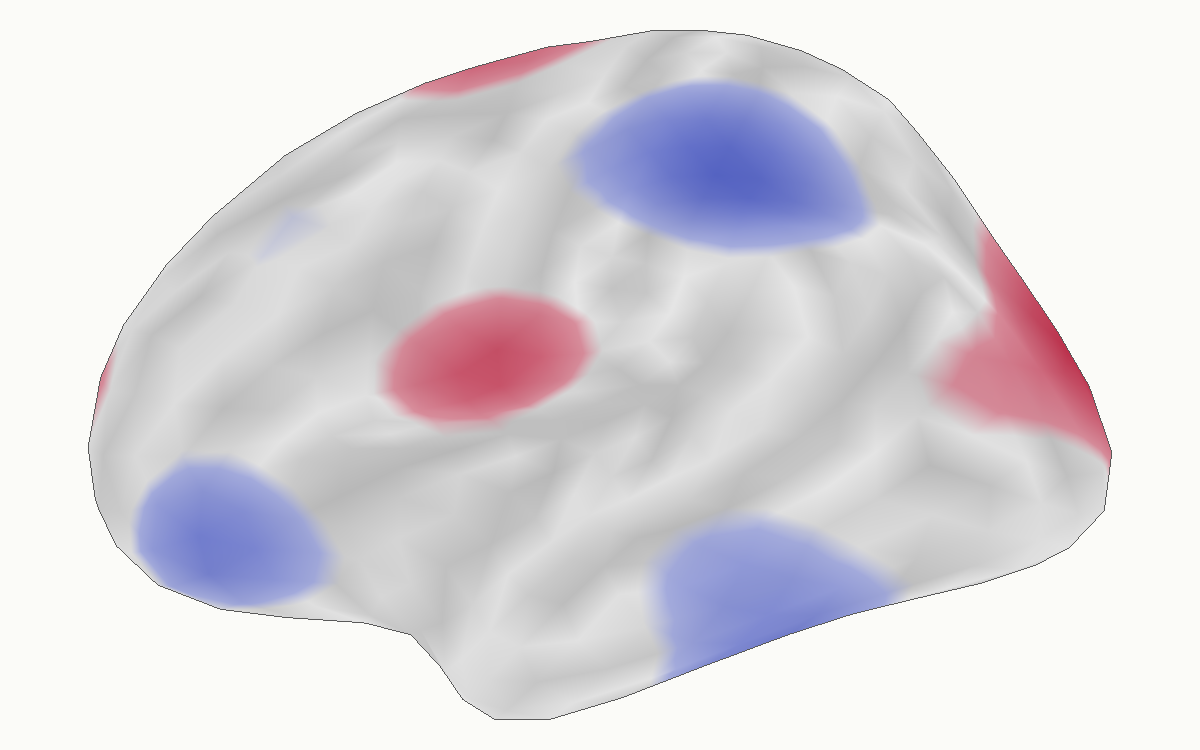
The colour at each covered sample comes from the barycentrically interpolated scalar. Thresholding and palette mapping happen after that interpolation, so a threshold boundary may cross a triangle instead of jumping from one mesh edge to the next. The per-sample z-buffer independently decides which folded surface fragment is visible.
Surfaces are lit with interpolated normals, so cortical form reads
without drawn outlines, and the overlay keeps its colour-bar colour to
within a few percent at any viewing angle. Coarse meshes are subdivided
for display (subdivide = "auto"); the subdivision keeps
every original vertex value and never creates a suprathreshold region
that linear interpolation would not.
How do you build the four canonical views?
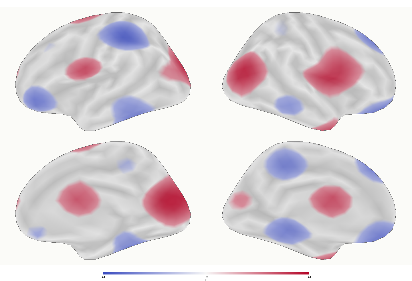
surface_figure() renders every hemisphere-view
combination with one shared threshold, colour limit, and palette, and
adds the colour bar. The camera contract is explicit:
camera_mode = "canonical" is strict orthographic output,
while "presentation" adds a small declared obliquity.
figure <- surface_figure(
  lh = inflated$lh,
  rh = inflated$rh,
  values = list(lh = stat_lh, rh = stat_rh),
  anatomy = sulc,
  views = c("lateral", "medial"),
  threshold = 1.5,
  tail = "two_sided",
  limits = c(-4, 4),
  alpha_ramp = 0.25,
  legend_title = "z",
  panel_width = 720,
  panel_height = 450,
  antialias = 3
)
In an interactive session, plot(figure) draws the same
figure on the current graphics device.
The lateral and medial views differ because each panel rasterizes the
visible triangles from its own camera. Internal sulcal occlusion edges
are not drawn as outlines; outer_contour = TRUE marks only
cortex touching background that is connected to the image exterior.
Where do the medial wall and cortex mask enter?
A real cortex mask is a separate domain object, not a consequence of
parcel value zero. Pass one logical value per vertex. Triangles touching
a masked vertex never receive overlay colour. medial_wall
then controls whether the excluded domain is quietly shaded, omitted, or
outlined.
masked_panel <- render_surface_rgba(
  inflated$lh,
  vertex_values = statistic,
  anatomy_metric = sulcal_depth,
  cortex_mask = cortex_label,
  medial_wall = "shade"
)
Atlas-aware callers such as neuroatlas::plot_brain() can
resolve the mask and its provenance from an atlas annotation. At this
low level, neurosurf requires the caller to supply the domain explicitly
rather than guessing that an atlas label of zero always means medial
wall.
How should a volume become a continuous surface field?
For a continuous image, use explicit trilinear sampling through the cortical ribbon. Interpolation, aggregation across depth, and tangential smoothing are separate choices. The following contract samples five depths, averages them, and applies no surface smoothing:
mapped <- vol_to_surf(
  white_surface,
  pial_surface,
  statistic_volume,
  fun = "avg",
  sampling = "thickness",
  interpolation = "linear",
  depth = seq(0.1, 0.9, length.out = 5),
  aggregate = "mean",
  surface_smooth_fwhm = 0
)
Use interpolation = "nearest" for discrete nearest-voxel
sampling. The historical Gaussian KNN behaviour remains available as
interpolation = "legacy". Categorical
aggregate = "mode" is intentionally incompatible with
linear interpolation.
Which function draws what you need?
Start from the output you want:
| You want | Use |
|---|---|
| A finished static figure: views by hemispheres, one colour bar |
surface_figure(), then
write_surface_figure()
|
| One panel, or a custom layout you compose yourself |
render_surface_rgba(), then
write_surface_rgba()
|
| To rotate and inspect a map on your desktop during analysis | view_surface() |
| An interactive figure in an HTML report or Shiny app |
surfwidget(); see
vignette("interactive-surfaces")
|
The first two run headlessly and deterministically; the last two are interactive sessions.
Two companion packages build on the same renderer, so their pixels
agree with the figures here: neuroatlas::plot_brain() adds
atlas-derived masks, labels, and orientation marks, and
neuromosaic::surf_montage() starts from a statistic volume
rather than vertex values. Atlas outlines belong in figures about the
atlas; the default continuous-statistic view omits them.
Next steps
-
vignette("displaying-surfaces")covers lower-level RGL rendering and local snapshots. -
vignette("interactive-surfaces")builds interactive bilateral report widgets withsurfwidget(). -
vignette("introduction-to-neurosurf")introducesSurfaceGeometry,NeuroSurface, and related data structures. -
?surface_figure,?render_surface_rgba,?surface_threshold_segments, and?vol_to_surfdocument the complete computational contracts.